
The eyelids play an important role in protecting the globe and the adnexal structures are crucial in producing clear vision.
The skin covering the eyelids is the thinnest on the body, measuring approximately 0.5 mm thick and lacking subcutaneous fat. The eyelids serve multiple functions:
The eyelids also contain meibomian glands which provide the lipid component of tear film, prevent the eyes from drying by blinking.
The inner structure of the eyelids is supported by tarsal plates. These are fibrous layers which gives the eyelids its shape and serve as attachment points for the eyelid muscles.
There are two tarsal plates: one on the upper eyelid called the superior tarsus and one on the lower eyelid called the inferior tarsus.
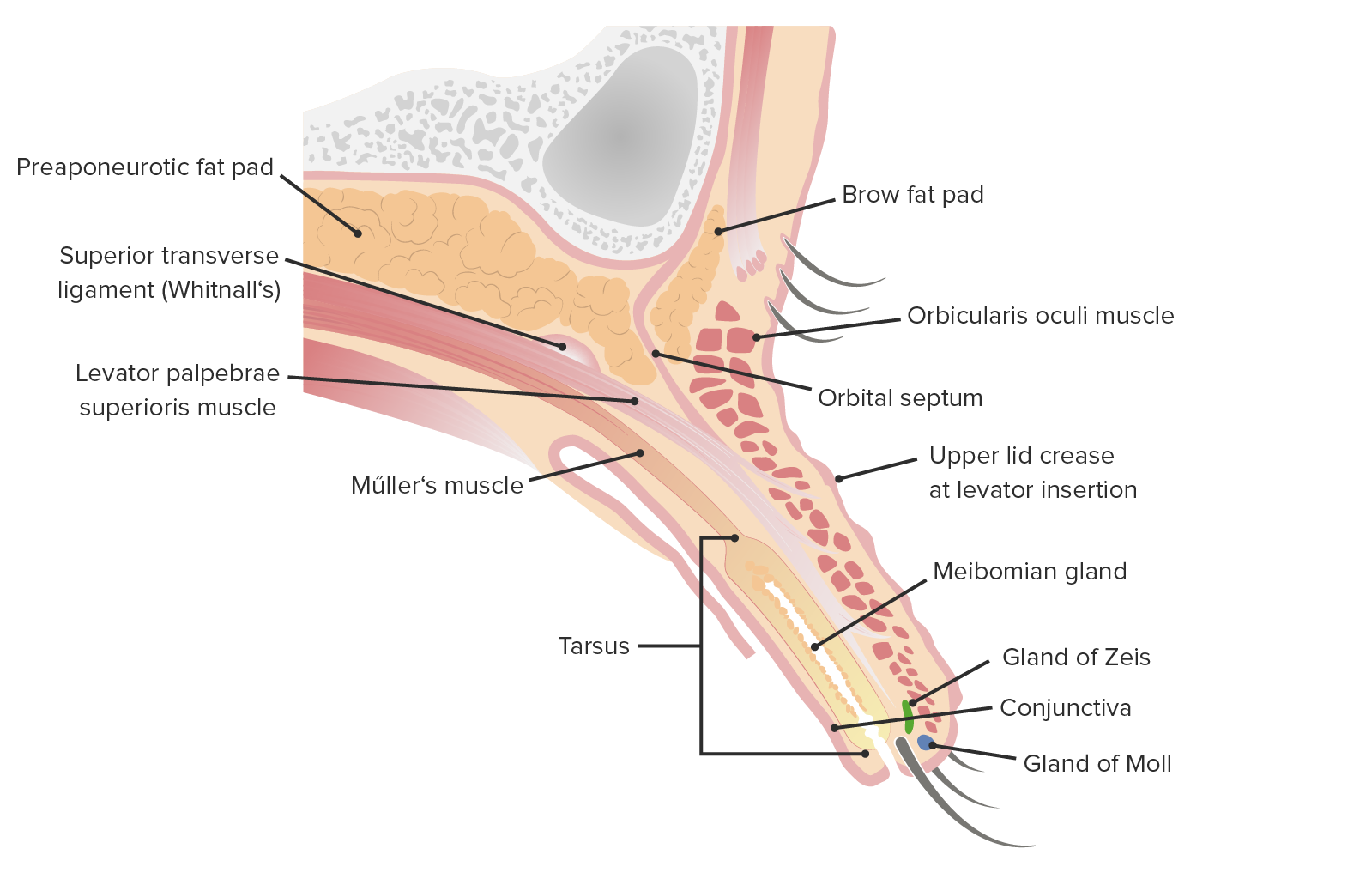
Diagram showing the superior tarsus and associated adnexal structures.
At the medial end of each tarsus, the medial palpebral ligament is formed (see diagram). This ligament crosses over the lacrimal sac (which we will discuss later), to attach to the maxilla (which we will also discuss later).
The medial palpebral ligament is also sometimes called the tendo oculi and is around 4 mm in length and 2 mm in width.

Diagram showing structures associated with the eyelids. Image courtesy of Lecturio
The eyelashes contain two types of glands:
The secretions produced by these two glands protect the surface of the eyelid.